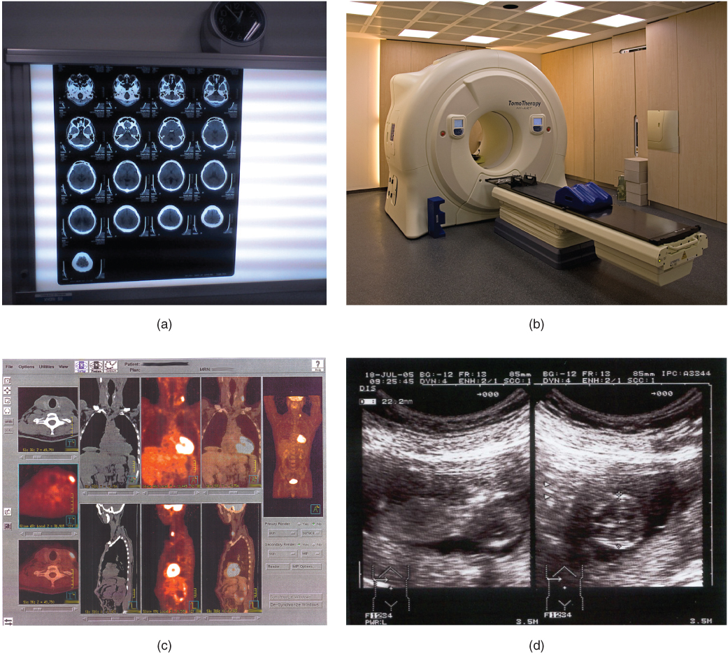
Sectional X-rays Of One Layer At A Time
HVL is an important quality control test as it is used to measure whether or not there. The HVL of an x-ray beam is defined as the amount of absorbing material that is needed to reduce the beam to half of its original potential.

Adenoid And Palatine Tonsil On Lateral Radiograph Radiology Case Radiopaedia Org Radiology Radiographer Palatine
The development of optical coherence tomography OCT has changed that.

Sectional x-rays of one layer at a time. The intensity of an x-ray beam at a given point number of photons per cross -sectional area per unit exposure time depends on the distance of the measuring device from the focal spot. Identify one metamorphic rock that could have been formed by the contact metamorphism within rock unit 1. Additionally the setup can be used to identify.
If your license expiration date is within 2 weeks of submitting your answers fax a copy to 502 327-7921. It features stars being torn apart by black holes galactic collisions and novae and neutron stars that build up layers of plasma that then explode into space. The growth rate of W and Ni layers were calibrated to be 18.
HVL is an indirect measure of photon energy or beam hardness. The half value layer expresses the thickness of absorbing material needed for reduction of the incident radiation intensity by a factor of two. The sequence of rock types found in the walls of the Grand Canyon are shown.
Computed tomography CT A radiographic method of viewing a three-dimensional image of a layer of body structures which is constructed by a computer from a series of plane cross-sectional X-ray images made along an axis. The names of rock formations are shown and the upper and lower boundaries of each formation are indicated by dashed lines. A sheet of lead foil surrounds all the films inside the box to protect them from damage by stray radiation or chemical fumes during storage.
Sectional x-rays of one layer at a time. In radiography technologists use the half value layer HVL to measure the quality or intensity of the beam. With this knowledge the thickness of different samples is being determined.
The growth of the thickness of alternate layers of W and Ni was controlled by interrupting the sputtered flux by a manually driven shutter which was operated after prefixed time duration. Certificate Issuance Your certificate will be scored the same day or next business day. The half value layer decreases as the atomic number of the absorber increases.
Periapical x-ray machines are typically mounted on the wall inside each treatment room. Until recently the cross sectional examination of paintings involved the microscopic examination of physical samples taken directly from the painting. The cycle is repeated until the desired thickness is reached say 100 atomic.
For a given beam the intensity is inversely proportional to the square of the distance from the source. DIDbase I0e basex1 sinW 1 sin2 W dx 7 Integration over the examined area results in. O The travel time for a reflected ray is given by.
Time captured on small film cards inserted in the mouth. OCT technique uses low coherence light and Michelson interferometry to examine thin semitransparent structures. Relating to the stomach.
T REFACTED xV 2 2H 1 V 2 2 V 1 2 12 V 1 V. The rock layers have not been overturned. Fluoroscopy time of 67 seconds is well less that the overall average of 147 seconds.
Inflammation of the stomach and small intestine. Find the magnitude of the electric field strength in the ring. This procedure was performed entirely using low dose settings 3.
Layer thickness determination via the absorption of X-rays Max-Planck-Institut fur extraterrestrische Physik Giessenbachstraße 85748 Garching October 25 2007 Aim of the experiment. Allow up to 10 days turnaround time. X-Ray interaction with matter and attenuation.
T REFLECTION x 2 4H 1 2 12 V 1. The magnetic induction produced by the solenoid varies with time as B bt where b is a constant. A long solenoid of cross-sectional radius a has a thin insulated wire ring tightly put on its winding.
An X-ray laser device was proposed as part of the Reagan Administration s Strategic Defense Initiative in the 1980s but the only test of the device a sort of laser blaster or death ray powered by a thermonuclear explosion gave inconclusive results. Inflammation of the pancreas. The radiographic film is composed of a base and an emulsion layer joined together by the substratum.
Bones attenuate most lungs attenuate least and blood vessels are in between. One half of the ring has the resistance η times that of the other half. First a single layer coating of a linear attenuation coe cient base is considered named as base coating tested by GI-XRD.
For example 35 m of air is needed to reduce the intensity of a 100 keV X-ray beam by a factor of two. O This is the equation for a hyperbola where H 1 is the layer thickness. IDbase I0 2 66 66 66 66 64 e.
Visual examination of the bronchi. This procedure was performed in the same room and same month as the higher than usual IVC filter placement REFERENCE 1. Consideration the fraction of the beam absorbed by each layer.
OCT is well known to the retinal specialist. Inflammation of the stomach. The emulsion may be coated on one side single emulsion film or both.
The X-rays are received by. You must score at least a. The images indicate the X-ray absorption called attenuation of tissues eg.
Consider solid A formed exclusively from copper and gold atoms in which two atomic layers of gold atoms are put down followed by two atomic layers of copper atoms followed by two atomic layers of gold atoms etc. Equation 3 now takes the form of Figure1b. Half Value Layer.
O The travel time for the refracted wave is given by. This is useful for studying the patients jaw and the position of the. With this experiment a basic understanding of the absorption of X-rays shall be achieved.
The reason for this decrease in intensity is that the x-ray beam spreads out. Please be sure to verify that we received your answer sheet. Panoramic pan x-rays generate a 5 x 11 or 15 cm x 30 cm wrap-around radiographic image of the patients mouth.
The time following birth relating to the baby. The time durations for shutter operation were decided after calibrating the growth rate during a test run which was carried out just before actual deposition. This suggests that the procedure was less difficult than average.
It is now possible to grow certain solids virtually an atomic layer at a time. One film per packet as for two films per packet. There are two main features of the half value layer.

Pin On Sandalwood Furniture

Ensembles Composables Et Chaises Longues Costco Fabric Sectional Sectional Top Grain Leather Sofa

Cindy Crawford Home Metropolis Way Steel Microfiber 3 Pc Sectional With Cuddler Curved Sofa Living Room Cindy Crawford Home Home
Pericardial Effusion Cardiac Tamponade Chapter 25 Emergency Cross Sectional Radiology

7pc Wicker Patio Furniture Sectional Sofa Set With Cushions Orange Wicker Patio Furniture Sectional Patio Furniture Sofa Set

Pin On Living Room Ideas

Park Sectional Sofa Sectional Sofa Chic Sofa Quality Mattress

How To Place A Rug Under A Sectional Sofa Living Room Rug Placement Sectional Sofas Living Room Area Rug Placement Living Room

Cervical Mri A Systematic Reading Vertebrae Alignment Discs Cervical Medullary Cord The T2 Sagittal And Axials Are Mri Cervical Human Skeleton Anatomy
Pin On D R E A M H O M E

Pin On 3d Models Of Furniture

Pin On Garden
Medical Imaging Anatomy And Physiology
Thoracic Aortic Aneurysm And Rupture Chapter 21 Emergency Cross Sectional Radiology
![]()
Radiological Anatomy X Ray Ct Mri Kenhub

Beste Von Couchgarnitur Gebraucht Kaufen Set Teras Set Ruang Keluarga Set Sofa

Hand Bone X Ray X Ray Hand Bone Medical

What Is A Sinus Rentention Cyst A Maxillary Sinus Retention Cyst Do I Need Sinus Surgery For Retention Cysts Mucocel Sinus Surgery Sinusitis Maxillary Sinus

Furniture Home Furnishings Find Your Inspiration Canape D Angle Modulaire Canape 4 Places Canape Modulable
Post a Comment for "Sectional X-rays Of One Layer At A Time"